Cytokinesis is one of the most significant steps that occurs during the last phase of cell division. Cytokinesis means the division of the cytoplasm of parental cells into two daughter cells. There are distinct features present in cytokinesis in animal cells and in plant cells. The partitioning of cytoplasm during meiosis and related sexual reproduction also determines the fate of the resulting daughter cells.
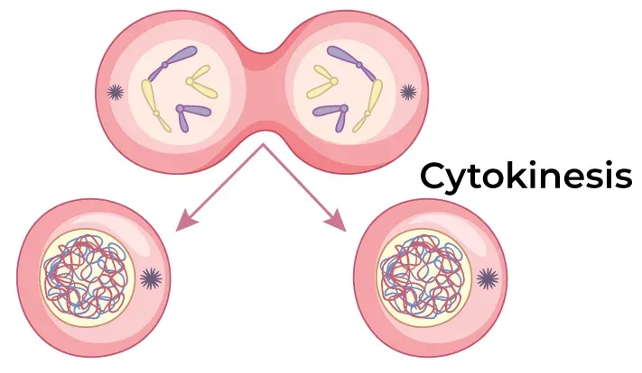
Mitosis accomplishes not only the segregation of duplicated chromosomes into two daughter nuclei (karyokinesis), but the cell itself is divided into two daughter cells by the separation of cytoplasm, called cytokinesis, at the end of which cell division is completed. After the process of karyokinesis in telophase, two nuclei are formed in a cell. Now, we have to divide this nucleus into a complete cell, and the process of cytokinesis starts from here.
Cytokinesis in Animal Cells
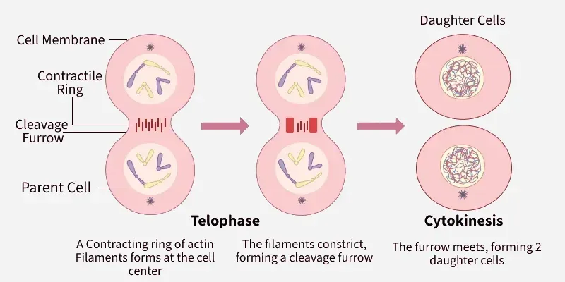
In animal cells, cytokinesis occurs by the formation of a cleavage furrow. After nuclear division, a contractile ring made of actin and myosin forms beneath the plasma membrane at the equatorial region. This ring contracts, deepening the cleavage furrow until the cytoplasm divides, resulting in two separate daughter cells.
It has the following stages:
- Formation of the Contractile Ring: In animal cells, the first step is the formation of a contractile ring composed of actin and myosin filaments. Its positioning is guided by central spindle microtubules, playing a crucial role in centring the actomyosin contractile ring within the cell.
- Cleavage Furrow Regression: As a result of the movement of actin and myosin filaments, a cleavage furrow is formed, which later leads to the construction of a deep cleavage furrow.
- Formation of Midbody: An intracellular bridge is formed between the two separating daughter cells with a specialised transient structure, which is called the midbody. This mid-body is made up of tightly bundled anti-parallel microtubules that connect these two daughter cells at the end of cytokinesis in animal cells.
- Abscission: The last phase is abscission, which produces two daughter cells. Numerous protein complexes required for transport, such as ESCRT-3 (endosomal sorting complex), aid in positioning and abscission. These filaments surround the mid-body and contribute to the abscission process. So, the cell division ends with the cutting of microtubules, and this intracellular bridge unites the two daughter cells in exact positions designated by the mid-body.
This complete process of cytokinesis in an animal cell can be summarised in 4 phases. These are:
Phases | Explanation |
|---|---|
Initiation | The contractile ring initiates and starts to build a cleavage furrow. This happens in the anaphase. |
Contraction | As telophase starts and anaphase ends, the contractile ring keeps contracting and widening the cleavage furrow. |
Membrane insertion | The process of inserting a newly produced cell membrane between two newly forming cells is known as membrane insertion. |
Completion | The contractile ring closes and divides the two new cells from one another at the point of completion. |
Cytokinesis in Plant Cells
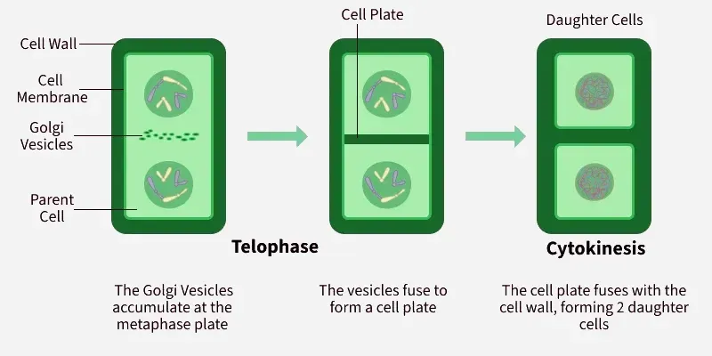
In plant cells, cytokinesis occurs by the formation of a cell plate due to the presence of a rigid cell wall. Vesicles from the Golgi apparatus accumulate at the centre of the cell and fuse to form the cell plate. The cell plate gradually grows outward and fuses with the existing cell walls, resulting in the formation of two separate daughter cells.
It has the following stages:
- Phragmoplast Formation: Between two chromosomes, a rearrangement of microtubules occurs, which leads to the formation of a microtubule-containing structure, phragmoplast, which guides the production of a new cell wall.
- Initiation of Cell plate formation: The cell organelle Golgi body, is responsible for bringing cell wall materials near the centre of the metaphase plate.
- Cell plate formation: The Golgi vesicles all fuse to form a continuous membranous structure called the cell plate.
- Cell plate expansion: The cell plate starts expanding towards the outer area with the help of microtubules.
- Fusion with the cell membrane: The cell plate then finally fuses with the cell membrane, thus resulting in the division of the cell. After the cell plate divides the cell, the plasma membrane closes, separating the two daughter cells. Trapped endoplasmic reticulum forms the plasmodesmata, or space, between the two cells, allowing chemicals to move from one to the other and signalling between cells.
Regulation of Cytokinesis
There are multiple ways in which cytokinesis is regulated. Some of these are:
- By Protein Kinases: Multiple mitotic protein kinases are involved in the regulation of cytokinesis. CDKs, or cyclin-dependent kinase, Polo Kinase (Plk1) and Aurora B kinase complex are a few examples of protein kinases. CDK does not allow cytokinesis to take place until anaphase by phosphorylating cytokinesis components. Plk1 and Aurora B kinase positively regulate the cytokinesis mechanism and are active while CDK1 becomes inactive.
- By Tyrosine Kinases: Some receptor tyrosine kinases help in regulating cytokinesis, regulating key signalling pathways that are involved in cell division. Some of these kinases help in activating downstream cascade reactions that affect the activity of cell cycle regulators.
- By Lipids: Lipids such as sphingolipids and phospholipids are the main constituents of the cell membrane. These lipids are a big part of the membrane remodelling process, such as membrane curvature generation and membrane scission, that eventually result in the cleavage formation, which further leads to cytokinesis. The lipids also act as signalling molecules, as phosphatidylinositol 4,5-bisphosphate helps in recruiting proteins that are important for cytokinesis.
Disorders and Abnormalities in Cytokinesis
The following points highlight the abnormalities seen during cytokinesis:
- Abscission, the final phase of cytokinesis, is the separation of cytoplasmic components from one another. Failure at this stage may result in cleavage furrow regression or the establishment of a permanent link between the two daughter cells.
- Cytokine dysregulation causes a variety of human ailments, including Lowe syndrome, blood disorders, cancer and female infertility. Cytokinesis failure can cause centrosome amplification, tetraploid cells, genetic instability, and apoptosis.
- Tetraploid cells are caused by cytokinesis failure, which occurs when diploid, mononucleated cells undergo mitosis but do not form a contractile ring.
- Anaphase spindle elongation does not occur in cells, possibly due to defective actin cytoskeleton reorganisation.
- Cytokinesis failure occurs in cells with mutations in the APC (Adenomatous Polyposis Coli) tumour suppressor. Some APC mutations may cause cytokinesis failure by interfering with the microtubule-dependent anchoring of the mitotic spindle.
- Myosin II is the primary motor protein necessary for cytokinesis. Since myosin motor activity is essential for furrow ingression, disrupting myosin localisation or activity may result in cytokinesis failure.
Significance of Cytokinesis
The following points help in understanding the importance of cytokinesis:
- Formation of daughter cells: The splitting of the original parent cell into daughter cells occurs with the help of cytokinesis.
- Maintenance of cell size: Cytokinesis helps in the proper distribution of cellular components between the daughter cells.
- Genetic stability: Any kind of irregularity seen during cytokinesis leads to genetic instability.
- Tissue growth and repair: In case of any damage to the cells, cytokinesis helps in the repair process by forming new cells.
- Development and Differentiation: Cytokinesis helps in the formation of special tissues, which thereby help in cell development and differentiation.
- Cellular Homeostasis: A balance is maintained in the body as cytokinesis maintains a proper order between cell growth and cell death.